Mob/ Whatsapp/Wechat: +86 138 806 76215 | Email: [email protected] / [email protected]
 Animal Tissue DNA Isolation kit is designed for rapid purification of total DNA (e.g., genomic, mitochondrial and pathogen) from a variety of sample sources including fresh or frozen animal tissues and cells, blood, or bacteria. The purified DNA is free of contaminants and enzyme inhibitors and is highly suited for PCR, Southern blotting, RAPD, AFLP and RFLP applications.
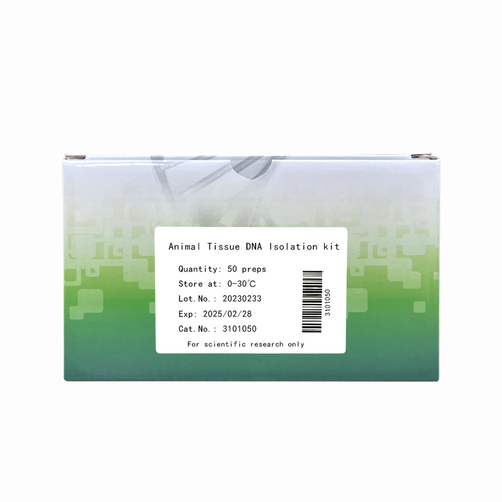
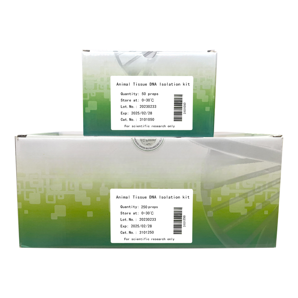
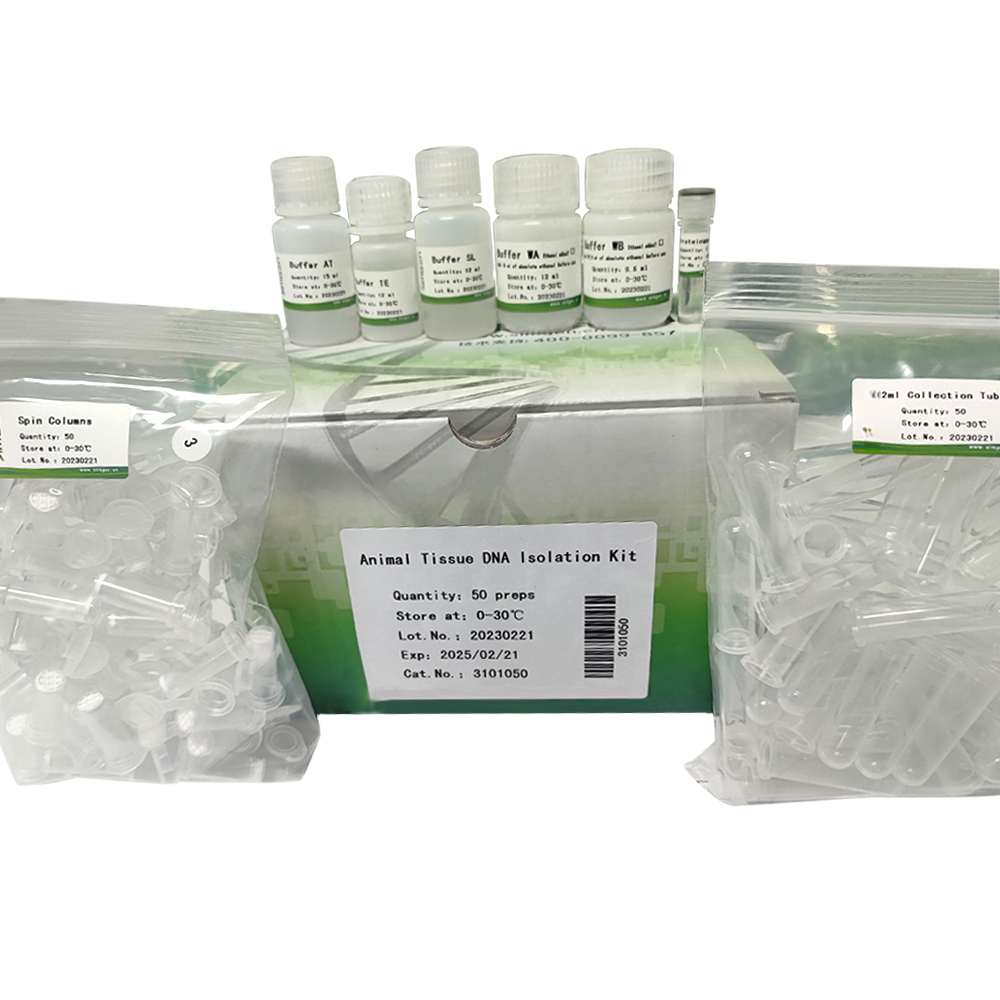
 | Animal Tissue DNA Isolation kit | 5 preps | 50 preps | 250 preps |
| Cat. No. | 3101005 | 3101050 | 3101250 |
| Spin Columns | 5 pcs | 50 pcs | 250 pcs |
| 2 ml Collection Tubes | 5 pcs | 50 pcs | 250 pcs |
| Proteinase K | 120 µl | 1.2 ml | 1.2 ml×5 |
| Buffer AT | 1.5 ml | 15 ml | 75 ml |
| Buffer SL | 1.2 ml | 12 ml | 60 ml |
| Buffer WA | 1.9 ml | 12 ml | 56 ml |
| Buffer WB | 1.5 ml | 9.5 ml | 50 ml |
| Buffer TE | 1.2 ml | 12 ml | 60 ml |
| Manual | 1 copy | 1 copy | 1 copy |
1. Proteinase K can be transported at room temperature, please store at -20°C after receiving the product.
2. All other reagents can be stored at room temperature for up to 2 years without showing any reduction in performance, and would be stable more than 2 years if stored at 2-8℃.
3. During shipment or storage in cool ambient conditions, precipitates may form in Buffer AT. Dissolve such deposits by warming the solution at 70℃ and gently shaking.
Animal Tissue DNA Isolation kit is designed for rapid purification of total DNA (e.g., genomic, mitochondrial and pathogen) from a variety of sample sources including fresh or frozen animal tissues and cells, blood, or bacteria. The purified DNA is free of contaminants and enzyme inhibitors and is highly suited for PCR, Southern blotting, RAPD, AFLP and RFLP applications. The buffer system is optimized to allow direct cell lysis followed by selective binding of DNA to the column membrane. After lysis, the DNA isolation procedure can be completed in as little as 20 minutes. Simple centrifugation processing completely removes contaminants and enzyme inhibitors, such as proteins and divalent cations. Purified DNA is eluted in Buffer TE or water, ready for use in downstream applications. The purified DNA typically has an A260/A280 ratio between 1.7 and 1.9, and the DNA fragments are from 100 bp to 50 kb in size, with of 30 kb predominating.
1. ethanol (96–100%)
2. 1.5 ml microcentrifuge tubes and pipet tips (pipet tips with aerosol barriers for preventing cross-contamination are recommended)
3. Microcentrifuge(s) (with rotor for 1.5 ml and 2 ml microcentrifuge tubes)
4. Thermostat water bath or Thermomixer with 2 ml inlays
5. Vortexer
6. 50 mg/ml RNase A (maybe needed)
7. PBS Buffer (maybe needed for formalin-fixed tissues)
8. Liquid nitrogen and mortar (maybe needed)
1. Prepare a thermomixer with 2 ml inlays or a water bath at 56℃ and 70℃ (37℃ should be added if gram-positive bacteria DNA needs to be purified), incubate Buffer AT and Buffer TE at 56℃.
2. Add absolute ethanol to Buffer WA and Buffer WB according to the instructions on the label of the reagent bottle, and tick the box on the label to mark "Ethanol added".
Maximum Amounts of Starting Sample
Sample | Amount |
Animal tissue | 10~25 mg |
Mammalian blood, bone marrow | 200 µl |
Bird or fish blood (with nucleated erythrocytes) | 5~10 µl |
Mouse tail | 0.6–1.2 cm |
Rat tail | 0.6 cm |
Cultured cells | 200 µl,5-10×106 |
Bacteria | 2 ×10⁹ |
For blood with non-nucleated erythrocytes, such as mammals,Saliva, follow step 1a.
For blood with nucleated erythrocytes, such as birds, fish, or frogs, contains nucleated erythrocytes, follow step 1b.
For cultured cells, follow step 1c.
For gram-negative bacteria, follow step 1d.
1a. Non-nucleated: Pipet 20 µl Proteinase K into a 1.5 ml or 2 ml microcentrifuge tube (not provided). Add 200 µl anticoagulated blood. Continue with step 2.
* If RNA-free genomic DNA is required, add 4 µl RNase A (50 mg/ml) and incubate for 2 min at room temperature (15–25°C) before continuing with step 2.
1b. Nucleated: Pipet 20 µl Proteinase K into a 1.5 ml or 2 ml microcentrifuge tube (not provided). Add 5–10 µl anticoagulated blood. Adjust the volume to 220 µl with PBS buffer. Continue with step 2.
* If RNA-free genomic DNA is required, add 4 µl RNase A (50 mg/ml) and incubate for 2 min at room temperature before continuing with step 2.
1c. Cultured cells: Centrifuge the appropriate number of cells (maximum 5×10⁶) for 5 min at 300×g. Resuspend the pellet in 200 µl PBS buffer. Add 20 µl Proteinase K. Continue with step 2.
* When using a frozen cell pellet, allow cells to thaw before adding PBS until the pellet can be dislodged by gently flicking the tube. Ensure that an appropriate number of cells is used. For cell lines with a high degree of ploidy (e.g., HeLa cells), it is recommended to use less than the maximum number of cells listed in Table 1.
* If RNA-free genomic DNA is required, add 4 µl RNase A (50 mg/ml), mix by vortexing, and incubate for 2 min at room temperature before continuing with step 2.
1d. Collect 1-3 ml of bacterial culture with a 1.5 ml microcentrifuge tube, add 100 μl Buffer TE, vortex and shake to suspend the bacteria thoroughly. Add 100 μl lysozyme solution, mix well by vortexing for about 15 sec. Then incubate at 37 °C for 30 min. Add 20 μl Proteinase K. Continue with step 2.
* Most bacteria can be fully broken after incubated at 37°C for 30 minutes, but some bacteria with particularly thick cell walls (such as Staphylococcus aureus) can appropriately extend the incubate time to 1 hour.
* Some media containing divalent cations (such as MRS medium, etc.) will inhibit the activity of lysozyme, a washing step should be added after the bacteria are collected: add 1 ml of distilled water, vortex the suspended bacteria and centrifuge at 12,000 rpm for 30 seconds, discard the distilled water, add 200 μl of Buffer TE, vortex to fully suspend the bacteria.
* Lysozyme solution preparation method: 100 mg/ml lysozyme solution is prepared according to the ratio of 100 mg lysozyme per 1 ml of deionized pure water.
* Lysozyme solution will seriously reduce the efficiency after freezing and thawing, please use freshly prepared or freeze-thaw no more than once lysozyme solution.
2. Add 200 µl Buffer SL. Mix thoroughly by vortexing and incubate at 56°C for 10 minutes.
* It is essential that the sample and Buffer SL are mixed immediately and thoroughly by vortexing or pipetting to yield a homogeneous solution.
3. Add 200 µl ethanol (96–100%) to the sample and mix thoroughly by vortexing.
* It is important that the sample and the ethanol are mixed thoroughly to yield a homogeneous solution.
4. Pipet the mixture from step 3 into a spin column (the spin column is placed in a 2 ml collection tube). Close the lid, and centrifuge at 12,000 rpm for 30 seconds.
* Avoid letting solution contact with edge of spin column, or it may affect purification.
5. Discard the filtrate. Place the spin column back to the 2 ml collection tube. Add 500 μl Buffer WA containing ethanol, close the lid and centrifuge at 12,000 rpm for 30 seconds.
* Ensure ethanol has been added into Buffer WA.
6. Discard the filtrate. Place the spin column back to the 2 ml collection tube. Add 600 μl Buffer WB containing ethanol, close the lid and centrifuge at 12,000 rpm for 30 seconds.
* Ensure ethanol has been added into Buffer WB.
7. Discard the filtrate. Place the spin column back to the 2 ml collection tube. Centrifuge at 14,000 rpm for 1 minute.
* If the top speed could not reach 14,000 rpm, centrifuge at top speed for 2 minutes.
* Do not omit this step, otherwise, it may cause problems in downstream applications due to the residual ethanol in the eluate.
8. Discard the 2 ml collection tube. Place the spin column to a new 1.5 ml centrifuge tube, Add 100-200 μl pre-warmed Buffer TE to the center of the membrane. Close the lid, incubate for 1 minute at room temperature, then centrifuge at 12,000 rpm for 30 seconds.
* Do not allow the spin column to dry, or it may affect DNA elution.
* Recovery may be increased by 10-20 % if incubate longer (5-15 minutes) after Buffer TE addition.
9. Discard the spin column. Eluted DNA can be used in downstream applications, or store at -20℃ for later use.
1. Cut up to 25 mg tissue (up to 10 mg spleen; for rodent tails, place one (rat) or two (mouse) 0.4-0.6 cm lengths of tail) into small pieces, and chop it into homogenate using scalpel, then transfer the tissue in a 1.5 ml microcentrifuge tube.
* We strongly recommend chopping the tissue into homogenate to enable more efficient lysis, especially connective tissue such as skin, tendon, and rodent tails, etc. If desired, lysis time can be reduced by grinding the sample in liquid nitrogen.
2. Add 20 μl proteinase K and 180 μl Buffer AT prewarmed at 56℃. Disperse the sample thoroughly by vortexing for a few seconds.
3. Incubate at 56℃ for 10~30 minutes. Vortex occasionally during incubation to lyse the sample completely, or place in a thermomixer, shaking water bath, or on a rocking platform.
* Lysis time varies depending on the type of tissue processed. Incubate longer until the tissue is completely lysed if there are still insoluble particulates present after 30 minutes’ incubation. If it is more convenient, samples can be lysed overnight; this will not affect them adversely.
* (Optional) RNase A treatment: If RNA-free genomic DNA is required, add 5 μl RNase A (50 mg/ml) after lysis, mix by vortexing for 30 seconds, and incubate for 2 min at room temperature, then continue with step 4. RNA may inhibit some downstream enzymatic reactions, but it does not affect PCR-related downstream applications. Transcriptionally active tissues such as liver and kidney contain high levels of RNA, which will copurify with genomic DNA. For tissues that contain low levels of RNA, such as rodent tails, or if residual RNA is not a concern, RNase A digestion is not necessary.
4. Add 200 μl Buffer SL, mix by vortexing for 15 seconds. Incubate at 70℃ for 10 minutes.
5. Add 200 μl ethanol (96-100%), mix by vortexing for 30 seconds.
6. Pipet the mixture from step 5 into a spin column (the spin column is placed in a 2 ml collection tube). Close the lid, and centrifuge at. 12,000 rpm for 30 seconds.
* Avoid letting solution contact with edge of spin column, or it may affect purification.
7. Discard the filtrate. Place the spin column back into the 2 ml collection tube. Add 500 μl Buffer WA containing ethanol, close the lid and centrifuge at 12,000 rpm for 30 seconds.
* Ensure ethanol has been added into Buffer WA.
8. Discard the filtrate. Place the spin column back into the 2 ml collection tube. Add 600 μl Buffer WB containing absolute ethanol, close the lid and centrifuge at 12,000 rpm for 30 seconds.
* Ensure ethanol has been added into Buffer WB.
9. Discard the filtrate. Place the spin column back into the 2 ml collection tube. Centrifuge at 14,000 rpm for 1 minute.
* If the top speed could not reach 14,000 rpm, centrifuge at top speed for 2 minutes.
* Do not omit this step, otherwise, it may cause problems in downstream applications due to the residual ethanol in the eluate.
10. Discard the 2 ml collection tube. Place the spin column into a new 1.5 ml collection tube (not provided). Add 100 - 200 μl pre-warmed Buffer TE to the center of the membrane. Close the lid, incubate for 1 minute at room temperature, then centrifuge at 12,000 rpm for 30 seconds.
* Do not allow the spin column to dry, or it may affect DNA elution.
* DNA yield may be increased by 10 - 20 % if incubate longer (5 - 15 minutes) after Buffer TE addition.
11. Discard the spin column. Eluted DNA can be used in downstream applications, or store at -20℃ for later use.
For large arthropod (e.g. shrimp, crab), please follow step 1a;
For small arthropod less than 500 mg (e.g. insect), please follow step 1b.
1a. Dissect exoskeleton using scalpel, cut 50-80 mg tissue into small pieces, and chop it into homogenate, then transfer the tissue in a 1.5 ml microcentrifuge tube.
1b. Weigh 300~500 mg tissue (including exoskeleton), transfer into a mortar, grind the sample into powder quickly in liquid nitrogen. Then weigh 60-120 mg powder into a 1.5 ml microcentrifuge tube precooled in liquid nitrogen.
2. Add 20 μl proteinase K stock solution and 250 μl Buffer AT prewarmed at 56℃. Disperse the sample thoroughly by vortexing for a few seconds.
3. Incubate at 56℃ for 10~30 minutes. Vortex occasionally during incubation to lyse the sample completely, or place in a thermomixer, shaking water bath, or on a rocking platform.
* (Optional) RNase A treatment: If RNA-free genomic DNA is required, add 5 μl RNase A (50 mg/ml) after lysis, mix by vortexing for 30 seconds, and incubate for 2 min at room temperature, then continue with step 4. RNA may inhibit some downstream enzymatic reactions, but it does not affect PCR-related downstream applications. Transcriptionally active tissues such as liver and kidney contain high levels of RNA, which will copurify with genomic DNA. For tissues that contain low levels of RNA, such as rodent tails, or if residual RNA is not a concern, RNase A digestion is not necessary.
4. Add 250 μl Buffer SL, mix by vortexing for 15 seconds. Incubate at 70℃ for 10 minutes.
5. Centrifuge at 14,000 rpm for 1 minute. Transfer 400 μl supernate into a new 1.5 ml microcentrifuge tube.
*If the top speed could not reach 14,000 rpm, centrifuge at top speed for 2 minutes.
6. Add 200 μl ethanol (96 - 100%), mix by vortexing for 30 seconds.
7. Pipet the mixture from step 6 into a spin column (the spin column is placed in a 2 ml collection tube). Close the lid, and centrifuge at. 12,000 rpm for 30 seconds.
* Avoid letting solution contact with edge of spin column, or it may affect purification.
8. Discard the filtrate. Place the spin column back into the 2 ml collection tube. Add 500 μl Buffer WA containing ethanol, close the lid and centrifuge at 12,000 rpm for 30 seconds.
* Ensure ethanol has been added into Buffer WA.
9. Discard the filtrate. Place the spin column back into the 2 ml collection tube. Add 600 μl Buffer WB containing absolute ethanol, close the lid and centrifuge at 12,000 rpm for 30 seconds.
* Ensure ethanol has been added into Buffer WB.
10. Discard the filtrate. Place the spin column back into the 2 ml collection tube. Centrifuge at 14,000 rpm for 1 minute.
* If the top speed could not reach 14,000 rpm, centrifuge at top speed for 2 minutes.
* Do not omit this step, otherwise, it may cause problems in downstream applications due to the residual ethanol in the eluate.
11. Discard the 2 ml collection tube. Place the spin column into a new 1.5 ml collection tube (not provided). Add 100 - 200 μl pre-warmed Buffer TE to the center of the membrane. Close the lid, incubate for 1 minute at room temperature, then centrifuge at 12,000 rpm for 30 seconds.
* Do not allow the spin column to dry, or it may affect DNA elution.
* DNA yield may be increased by 10 - 20 % if incubate longer (5 - 15 minutes) after Buffer TE addition.
12. Discard the spin column. Eluted DNA can be used in downstream applications, or store at -20℃ for later use.
1. Cut up to 25 mg formalin-fixed tissue, wash twice in 1 ml PBS to remove the fixative, discard supernate carefully by pipetting. chop the tissue into homogenate using scalpel, then transfer it into a 1.5 ml microcentrifuge tube.
* Tissue more than 25 mg may results in insufficient digestion of protease K, which would clog the spin column.
2. Add 20 μl proteinase K and 180 μl Buffer AT prewarmed at 56℃. Disperse the sample thoroughly by vortexing for a few seconds.
3. Incubate at 56℃ for 10~30 minutes. Vortex occasionally during incubation to lyse the sample completely, or place in a thermomixer, shaking water bath, or on a rocking platform.
* Lysis time varies depending on the type of tissue processed. Incubate longer until the tissue is completely lysed, if there are still insoluble particulates present after 30 minutes’ incubation. If it is more convenient, samples can be lysed overnight and not affect them adversely.
4. Add 200 μl Buffer SL, mix by vortexing for 15 seconds. Incubate at 70℃ for 10 minutes.
* If insoluble particulates (as bones, hair) are still present after the end of this step, centrifuge at 12,000 rpm for 1 minute. Transfer the supernate to a new 1.5 ml microcentrifuge tube (not provided). Then continue with step 5.
5. Add 200 μl ethanol (96 - 100%), mix by vortexing for 30 seconds.
6. Pipet the mixture from step 5 into a spin column (the spin column is placed in a 2 ml collection tube). Close the lid, and centrifuge at. 12,000 rpm for 30 seconds.
* Avoid letting solution contact with edge of spin column, or it may affect purification.
7. Discard the filtrate. Place the spin column back into the 2 ml collection tube. Add 500 μl Buffer WA containing ethanol, close the lid and centrifuge at 12,000 rpm for 30 seconds.
* Ensure ethanol has been added into Buffer WA.
8. Discard the filtrate. Place the spin column back into the 2 ml collection tube. Add 600 μl Buffer WB containing absolute ethanol, close the lid and centrifuge at 12,000 rpm for 30 seconds.
* Ensure ethanol has been added into Buffer WB.
9. Discard the filtrate. Place the spin column back into the 2 ml collection tube. Centrifuge at 14,000 rpm for 1 minute.
* If the top speed could not reach 14,000 rpm, centrifuge at top speed for 2 minutes.
* Do not omit this step, otherwise, it may cause problems in downstream applications due to the residual ethanol in the eluate.
10. Discard the 2 ml collection tube. Place the spin column into a new 1.5 ml collection tube (not provided). Add 100 - 200 μl pre-warmed Buffer TE to the center of the membrane. Close the lid, incubate for 1 minute at room temperature, then centrifuge at 12,000 rpm for 30 seconds.
* Do not allow the spin column to dry, or it may affect DNA elution.
* DNA yield may be increased by 10 - 20 % if incubate longer (5 - 15 minutes) after Buffer TE addition.
11. Discard the spin column. Eluted DNA can be used in downstream applications, or store at -20℃ for later use.
For pure gram-negative bacteria culture (e.g. E. coli), please follow step 1a;
For gram-negative pathogens (e.g. Treponema Pallidum) in anticoagulated blood, please follow step 1b.
1a. Harvest cells from 1-3 ml bacterial culture in a microcentrifuge tube (not provided) by centrifuging for 30 seconds at 12,000 rpm. Discard supernatant. Suspend pellet in 180 μl Buffer AT prewarmed at 56℃. Disperse the sample thoroughly by vortexing for a few seconds.
* Suspend pellet immediately after addition of Buffer AT, otherwise lysis maybe insufficient.
1b. Pipet 100 μl anticoagulated blood in a 1.5 ml microcentrifuge tube (not provided), add 100 μl Buffer AT prewarmed at 56℃. Disperse the sample thoroughly by vortexing for a few seconds.
2. Add 20 μl proteinase K, mix by vortexing for a few seconds.
3. Incubate at 56℃ for 10 minutes. Vortex occasionally during incubation to lyse the sample completely, or place in a thermomixer, shaking water bath, or on a rocking platform.
* The lysate should be clear after lysis. Incubate for 30 minutes if the lysate seems to be turbid.
4. Add 200 μl Buffer SL, mix by vortexing for 15 seconds. Incubate at 70℃ for 10 minutes.
* (Optional) RNase A treatment: RNA would copurify with genomic DNA in very fresh sample, it would inhibit some downstream enzymatic reactions, although it does not affect PCR-related downstream applications. If RNA-free genomic DNA is required, add 5 μl RNase A (50 mg/ml), mix by vortexing for 30 seconds, and incubate for 2 min at room temperature, then continue with step 5.
5. Add 200 μl Buffer SL, mix by vortexing for 15 seconds. Incubate at 70℃ for 10 minutes.
6. Add 200 μl ethanol (96 - 100%), mix by vortexing for 30 seconds.
7. Pipet the mixture from step 6 into a spin column (the spin column is placed in a 2 ml collection tube). Close the lid, and centrifuge at. 12,000 rpm for 30 seconds.
* Avoid letting solution contact with edge of spin column, or it may affect purification.
8. Discard the filtrate. Place the spin column back into the 2 ml collection tube. Add 500 μl Buffer WA containing ethanol, close the lid and centrifuge at 12,000 rpm for 30 seconds.
* Ensure ethanol has been added into Buffer WA.
9. Discard the filtrate. Place the spin column back into the 2 ml collection tube. Add 600 μl Buffer WB containing absolute ethanol, close the lid and centrifuge at 12,000 rpm for 30 seconds.
* Ensure ethanol has been added into Buffer WB.
10. Discard the filtrate. Place the spin column back into the 2 ml collection tube. Centrifuge at 14,000 rpm for 1 minute.
* If the top speed could not reach 14,000 rpm, centrifuge at top speed for 2 minutes.
* Do not omit this step, otherwise, it may cause problems in downstream applications due to the residual ethanol in the eluate.
11. Discard the 2 ml collection tube. Place the spin column into a new 1.5 ml collection tube (not provided). Add 100~200 μl pre-warmed Buffer TE to the center of the membrane. Close the lid, incubate for 1 minute at room temperature, then centrifuge at 12,000 rpm for 30 seconds.
* Do not allow the spin column to dry, or it may affect DNA elution.
* DNA yield may be increased by 10~20 % if incubate longer (5~15 minutes) after Buffer TE addition.
12. Discard the spin column. Eluted DNA can be used in downstream applications, or store at -20℃ for later use.